Position: Research scientist
Office address: Laboratory of Environmental Toxicology , Chulabhorn Research Institute
Phone number: +662-553-8555 ext. 8232
email: 
Education:
| 1993 | Bachelor of Science (Food Science and Technology), ChiangMai University (CMU), Thailand |
| 1996 | Master of Applied Science (Food Science and Nutrition), University of Western Sydney, Hawskerbury, Australia |
| 2008 | Doctor of Technical Science in Environmental Toxicology, Technology and Management (ETTM), Asian Institute of Technology (AIT), Thailand |
My own website: http://www.puiock-gallery.com/myself/index.html
Research work website: The Application of Dielectrophoresis in Environmental Studieshttp://dep.puiock-gallery.com
Gallery website: The Puiock Gallery.com http://www.puiock-gallery.com
Past Project
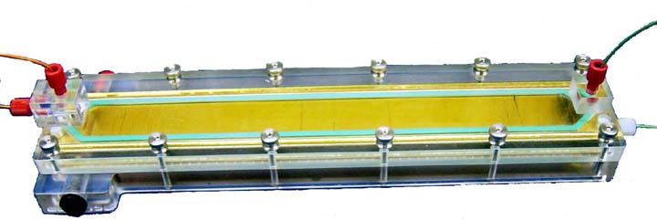
Dielectrophoresis Field-Flow Fractionation System for Detection of Aquatic Toxicants
Sittisak Pui-ock1, Mathuros Ruchirawat1,2 and Peter Gascoyne3
1Laboratory of Environmental Toxicology, Chulabhorn Research Institute, Bangkok, Thailand,
2Department of Pharmacology, Faculty of Science, Mahidol University, Bangkok, Thailand,
3Department of Molecular Pathology, M.D. Anderson Cancer Center, University of Texas, Houston, Texas 77030
Received for review May 29, 2008. Accepted July 11, 2008.
Abstract:
Dielectrophoretic field-flow fractionation (dFFF) was applied as a contact-free way to sense changes in the plasma membrane capacitances and conductivities of cultured human HL-60 cells in response to toxicant exposure. A micropatterned electrode imposed electric forces on cells in suspension in a parabolic flow profile as they moved through a thin chamber. Relative changes in the dFFF peak elution time, reflecting changes in cell membrane area and ion permeability, were measured as indices of response during the first 150 min of exposure to eight toxicants having different single or mixed modes of action (acrylonitrile, actinomycin D, carbon tetrachloride, endosulfan, N-nitroso-N-methylurea (NMU), paraquat dichloride, puromycin, and styrene oxide). The dFFF method was compared with the cell viability assay for all toxicants and with the mitochondrial potentiometric dye assay or DNA alkaline comet assay according to the mode of action of the specific agents. Except for low doses of nucleic acid-targeting agents (actinomycin D and NMU), the dFFF method detected all toxicants more sensitively than other assays, in some cases up to 105 times more sensitively than the viability approach. The results suggest the dFFF method merits additional study for possible applicability in toxicology.
For full detail of this paper, please visit
Pui-ock S, Ruchirawat M, Gascoyne P
Anal Chem80p7727-34(2008 Oct 15)
dFFF VDO clip
This VDO clip shows how dFFF can be applied for toxicity testing.
You can watch dFFF MV from youtube.com as shown below
Ongoing Project
Isolation of Circulating Tumor Cells by Dielectrophoretic Field-Flow Fractionation System
Circulating tumor cells (CTCs) that are shed from a primary cancer into peripheral blood and subsequently embed at a distal site where they subsequently grow into tumors are agents by which secondary disease occurs. Thus understanding the occurrence and properties of these cells is of great importance.
Current methods for CTC detection mostly use immunomagnetic enrichment of CTCs that can only detect tumors of epithelial origin; cell viability is not maintained, and most molecular information is lost. This study aims to develop a better separation of CTCs taken from blood by use of dielectrophoresis (DEP).
DEP is a contactless method which will not cause cell damages during separation. It separates cells by exposing cells in a flowing, low conductivity medium to a high frequency alternating electric field. Cells suspended in sucrose fluid will migrate to different levels within the fluid flow, depending on cell membrane capacitance. DEP system creates precisely controlled flow profiles that allow cell suspensions to be injected and withdrawn at very accurately determined positions in a microfluidic flow chamber.
Tumor cells flow through the chamber at different height from blood cells as a result of different cell membrane capacitance properties and size. Cancer cells are skimmed off and ready for cell culture, while the blood cells flow to waste.
Cell lysis is used to eliminate red blood cells before loading sample to DEP chamber. The study of mimicking CTC model by adding around 100 cells of breast, bile duct or liver cancer cell lines to 5 ml of normal blood shows that DEP isolated cancer cells could be re-cultured.
For poster presentation please go to thaiDEP website

